Cardiovascular disease remains the leading cause of mortality worldwide, yet its earliest stages develop silently over decades.1,2
For years, clinical medicine relied primarily on statistical risk estimators (such as the PREVENT equations or Pooled Cohort Equations) to calculate a patient’s probability of experiencing a heart attack or stroke over a 10-to-30-year horizon.1,3 While these population-level calculators provide a helpful baseline, they estimate statistical probability based on demographic averages—they cannot determine whether plaque is physically accumulating inside a specific patient's arteries.1
The Coronary Artery Calcium (CAC) scan serves as a vital complementary tool.1,4
By providing a direct, non-invasive anatomical view of the coronary vessels, a CAC scan refines clinical risk estimation by directly measuring the presence and extent of calcified atherosclerotic plaque.1,4
Here is an evidence-based guide to understanding CAC scans: how they work, how scores are categorized under contemporary guidelines, radiation safety, cost and insurance realities, how results guide medical therapy, and how a CAC scan compares to a Coronary CT Angiography (CCTA).
1. What Is a CAC Scan and What Does It Measure?
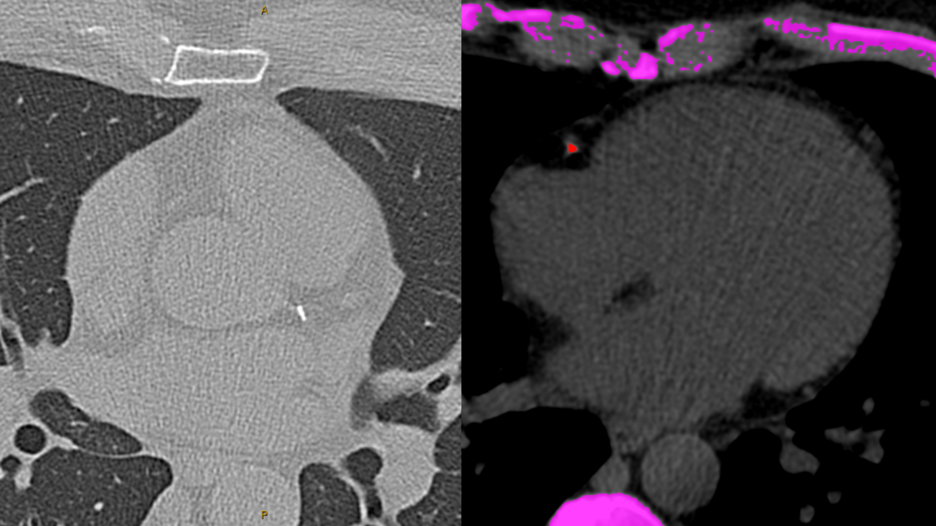
A Coronary Artery Calcium scan is a specialized, non-contrast computed tomography (CT) scan of the heart that takes roughly 10 to 15 seconds to complete.1,4
The scan specifically detects calcified atherosclerotic plaque within the walls of the coronary arteries:1,4
The Pathophysiology: Atherosclerosis begins when atherogenic particles (Apolipoprotein B / LDL particles) penetrate the endothelial lining of an artery, initiating local inflammation, macrophage recruitment, foam cell formation, and early non-calcified (soft) plaque accumulation.1,2
The Calcification Process: Over time, as part of the vascular remodeling and healing response to chronic inflammation, calcium hydroxyapatite crystals deposit within the plaque matrix.1,4
What Calcium Signifies: Calcification reliably indicates the physical presence of coronary atherosclerosis. If calcium is present in the coronary arteries, atherosclerotic disease is established.1,4
The total area and density of calcification are mathematically calculated into a standardized score known as the Agatston Score.4
2. Understanding Your CAC Score Categories
Under contemporary cardiology frameworks, including the 2026 ACC/AHA Multisociety Dyslipidemia Guidelines, CAC scores are classified across standardized categories to guide clinical risk stratification:1,4
Absent (CAC = 0 AU): No calcified plaque detected. This identifies an individual at very low near-term risk for cardiovascular events (typically < 1% annual event rate over the subsequent 5 to 10 years).1,5
Minimal Plaque (CAC = 1 to 9 AU): Detects very early subclinical calcification, confirming the presence of initial atherosclerotic disease.1
Mild Plaque (CAC = 10 to 99 AU): Confirms definite subclinical atherosclerosis and indicates higher risk than a score of zero, warranting lifestyle optimization and personalized risk evaluation.1,5
Moderate Plaque (CAC = 100 to 299 AU): Represents substantial atherosclerotic plaque burden. Under guideline recommendations, a score >= 100 AU (or >= 75th percentile for age and sex) strongly favors initiating or intensifying lipid-lowering therapy.1
Severe to Extensive Plaque (CAC >= 300 AU): Extensive coronary plaque burden. Patients with CAC >= 300 AU experience major adverse cardiovascular event (MACE) rates comparable to individuals with established clinical coronary artery disease (secondary prevention populations), warranting aggressive medical risk reduction.1,2
3. How a CAC Score Informs Medical Management
A CAC scan is not a replacement for comprehensive clinical assessment; it is a risk reclassifier that helps resolve therapeutic uncertainty when treatment decisions are borderline:1,3
1. Initiating or Deferring Lipid-Lowering Therapy
Under the 2026 ACC/AHA guidelines, CAC scoring serves as a primary tool to guide statin and lipid-lowering decisions in adults at borderline or intermediate calculated risk:1
When CAC = 0: Statin therapy can often be safely deferred or delayed, provided the patient does not have diabetes, is not a current cigarette smoker, does not have severe primary hypercholesterolemia (LDL-C >= 190 mg/dL), and does not have a strong family history of premature ASCVD.1
When CAC >= 100 AU (or >= 75th Percentile): Guidelines strongly recommend initiating lipid-lowering therapy (statins, ezetimibe, or PCSK9-targeted therapies) to lower Apolipoprotein B (ApoB) and slow cardiovascular disease progression.1
When CAC >= 300 AU: Indicates high risk, supporting intensive lipid lowering targeting lower ApoB and LDL-C thresholds.1
2. Primary Prevention Aspirin Decisions
The role of low-dose aspirin in primary prevention requires balancing cardiovascular protection against bleeding risks:1,6
Observational cohort studies and decision-analytic models demonstrate that for individuals with a CAC score >= 100 AU, low-dose aspirin provides a net clinical benefit only if the patient has a low bleeding risk and an estimated baseline ASCVD risk >= 5%.6
Conversely, in individuals with a CAC score of 0 or those with an elevated baseline bleeding risk, the risk of major bleeding outweighs potential cardiovascular benefits across all CAC strata.6 Professional society recommendations on aspirin for elevated CAC remain variable, making individualized clinical evaluation essential.1,6
3. Reinforcing Long-Term Adherence
Visual confirmation of coronary calcification has a proven impact on patient engagement. Clinical trials and registry data show that patients who review objective imaging of their coronary plaque demonstrate significantly higher long-term adherence to prescribed lifestyle modifications and preventive medications compared to those given statistical percentage estimates alone.1,4
4. CAC Scan vs. CCTA: What Is the Difference?
While both modalities utilize cardiac CT technology, a CAC scan and a Coronary Computed Tomography Angiography (CCTA) serve distinct clinical roles:1,2
Coronary Artery Calcium (CAC) Scan:
Coronary CT Angiography (CCTA):
Contrast Agent: Requires an intravenous (IV) iodinated contrast dye.
What It Measures: Full coronary visualization, including calcified plaque, non-calcified (soft) plaque, high-risk plaque characteristics (such as low-attenuation plaque or positive remodeling), and precise percentage of luminal stenosis.1,2
Radiation Exposure: Low to moderate, with real-world median doses around 7.4 mSv (though modern protocols can achieve lower exposure depending on scanner technology and heart rate control).7
Clinical Indication: Evaluating symptomatic patients (chest pain, unexplained dyspnea) or select individuals requiring detailed anatomical mapping of coronary stenosis and total plaque burden.1,2
5. Safety, Radiation, Cost, and Insurance
Understanding the practical details of a CAC scan helps patients make informed screening choices:
Radiation Exposure
A standard modern CAC scan delivers a median radiation dose of approximately 0.5 to 1.2 millisieverts (mSv).1,7 To put this in context:
The average individual receives roughly 3.0 mSv of background environmental radiation each year.
A CAC scan delivers less radiation than a standard diagnostic chest CT (~4–7 mSv) and is comparable to a routine screening mammogram (~0.4–0.7 mSv).1,7
For appropriately selected individuals, the diagnostic value of identifying subclinical cardiovascular disease outweighs the theoretical risks of low-dose radiation.1,4
Cost and Insurance Realities
Despite strong endorsement in clinical practice guidelines, commercial health insurers and Medicare generally do not cover CAC scans for asymptomatic screening.1
Because the procedure is non-contrast and takes only minutes, out-of-pocket pricing remains accessible. While hospital billing can reach $200 to $400, dedicated outpatient imaging centers typically charge between $50 and $250.1
At delaeMD, our telemedicine practice coordinates direct-access CAC scans for our patients through accredited imaging partners for approximately $100 out-of-pocket.
6. Who Should (and Should Not) Get a CAC Scan?
A CAC scan is a targeted diagnostic test intended for specific clinical scenarios:1,3
Appropriate Candidates:
Asymptomatic men aged >= 40 and women aged >= 45 with borderline or intermediate calculated 10-to-30-year cardiovascular risk where the decision to start statin therapy is uncertain.1
Individuals with a family history of premature ASCVD (heart attack in a first-degree male relative < 55 or female relative < 65).1
Individuals with elevated atherogenic particles (ApoB) or elevated Lipoprotein(a) [Lp(a)] where treatment thresholds benefit from anatomical risk reclassification.1
Who Does NOT Need a CAC Scan:
Younger Adults (< 35–40 years old): Plaque in younger individuals is predominantly non-calcified. A CAC score of 0 in a 28-year-old can provide false reassurance despite the presence of early soft plaque.1,2
Patients with Known Established CAD: Individuals with a history of myocardial infarction, coronary stenting, or bypass surgery already have confirmed advanced disease; a CAC scan will not alter their clinical management category.1
Very High-Risk Patients: Individuals with familial hypercholesterolemia (LDL-C >= 190 mg/dL) or diabetes already meet clear guideline indications for intensive lipid-lowering therapy regardless of their CAC score.1
The Bottom Line
Cardiovascular disease is largely preventable when identified and managed early.1,2 Rather than relying entirely on statistical calculations or waiting for clinical symptoms, a Coronary Artery Calcium scan provides objective anatomical evidence to refine individual risk.1,4
Knowing whether your score is 0, 50, or >= 300 provides clear, actionable guidance for you and your physician to optimize lipid targets, preventive medications, and lifelong cardiovascular health.1
References
Blumenthal RS, Morris PB, Gaudino M, et al. 2026 ACC/AHA/AACVPR/ABC/ACPM/ADA/AGS/APhA/ASPC/NLA/PCNA Guideline on the Management of Dyslipidemia: A Report of the American College of Cardiology/American Heart Association Joint Committee on Clinical Practice Guidelines. Journal of the American College of Cardiology. 2026;87(19):2624-2757.
van der Bijl P, Gulati M, Saraste A, et al. Contemporary, Non-Invasive Imaging Diagnosis of Chronic Coronary Artery Disease. The Lancet. 2025;406(10519):2577-2587.
Khan SS, Matsushita K, Sang Y, et al. Development and Validation of the American Heart Association's PREVENT Equations. Circulation. 2024;149(6):430-449.
Greenland P, Blaha MJ, Budoff MJ, Erbel R, Watson KE. Coronary Calcium Score and Cardiovascular Risk. Journal of the American College of Cardiology. 2018;72(4):434-447.
Budoff MJ, Young R, Burke G, et al. Ten-year association of coronary artery calcium with atherosclerotic cardiovascular disease (ASCVD) events: the multi-ethnic study of atherosclerosis (MESA). European Heart Journal. 2018;39(25):2401-2408.
Ajufo E, Ayers CR, Vigen R, et al. Value of Coronary Artery Calcium Scanning in Association With the Net Benefit of Aspirin in Primary Prevention of Atherosclerotic Cardiovascular Disease. JAMA Cardiology. 2021;6(2):179-187.
Einstein AJ, Williams MC, Weir-McCall JR, et al. Worldwide Radiation Dose in Coronary Artery Disease Diagnostic Imaging. JAMA. 2026;2845479.
Clinical Oversight & Authorship
Physician revieweddelaeMD Clinical Team
Clinical content at delaeMD is written and reviewed by licensed physicians, grounded in peer-reviewed evidence and current clinical guidelines, and intended for education — not as a substitute for individualized medical advice. Reviews are non-sponsored, physician-directed, and conducted independently of industry funding.